Myocardial deformation evaluated by two-dimensional echocardiography in lupic patients from a national hospital
DOI:
https://doi.org/10.47487/apcyccv.v1i3.66Keywords:
lupus erythematosus, echocardiography, Myocardial deformation, cardiomyopathiesAbstract
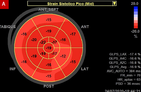
Background. Patients with systemic lupus erythematosus (SLE) are at high risk of cardiac compromise with high mortality. The subclinical diagnosis may improve their survival. Longitudinal myocardial deformation (strain) has been found to be useful in evaluating cardiac function in these patients.
Objectives. Our aims were to evaluate myocardial function by analyzing the two-dimensional (2D) global longitudinal strain, to compare the longitudinal strain in SLE patients with controls, and to determine the correlation with SLE activity index.
Material and Methods. 44 patients with SLE (50.0 ± 13 years) and 50 controls (49 ± 12 years) matched by age and sex, underwent transthoracic echocardiogram. Longitudinal strain was assessed using the speckle tracking method and SLE activity was estimated using the Systemic Lupus Erythematous Disease Activity Index (SLEDAI). A score of 4 or more, was defined as active SLE.
Results. 2D global longitudinal strain was lower in patients with SLE than controls (- 17.3% ± 1.9% vs. -20%, ± 1.9% p = 0.00). The left ventricular ejection fraction (LVEF) had no specific differences in both groups in 2D (p = 0.650) or three-dimensional (3D) (p = 0.718). In lupus patients, SLEDAI ranged from 0 to 10, and 63.8% were inactive. Negative correlations were found between the SLEDAI score and 2D LVEF (Pearson's r = -0.372, p = 0.017); no correlations were found between the SLEDAI score and the 2D global longitudinal strain (Spearman's rho = - 0.091 p = 0.582).
Conclusions: 2D global longitudinal strain was found to be decreased in the SLE group. This technique might can be a useful tool to assess cardiac function in these patiens.
Downloads
References
Villa-Forte A, Mandell B. Trastornos cardiovasculares y enfermedad reumática. Rev Esp Cardiol. 2011;64(9):809–17. Disponible en DOI: 10.1016/j.recesp.2011.05.009.
Gómez-León A, Amezcua-Guerra L. Manifestaciones cardiovasculares en el lupus eritematoso generalizado. Arch de Cardiologia de Mexico. Vol. 78 Número 4/Octubre-Diciembre 2008:421-30. Disponible en ISSN: 1665-1731.
Tsokos. Systemic lupus erythematosus. N Engl J Med. 2011;365(22):2110-21. Disponible en: DOI: 10.1056/NEJMra1100359.
Di Minno M, Forteb F, Antonel, et al. Speckle tracking echocardiography in patients with systemic lupus erythematosus: A meta-analysis. Eur J Intern Med. 2020 Mar;73:16-22. Disponible en DOI: 10.1016/j.ejim.2019.12.033.
Saad A, Cintora F, Pinasco D, et al. Evaluación de la función del ventrículo izquierdo en pacientes con lupus eritematoso sistémico mediante ecocardiografía tridimensional. Revista Argentina de Cardiología: 85(6); Diciembre 2017. Disponible den DOI: 10.7775/rac.es.v85.i6.12260.
Lang R, Badano L, Mor-Avi V, et al. Recommendations for Cardiac Chamber Quantification by Echocardiography in Adults: An Update from the American Society of Echocardiography and the European Association of Cardiovascular Imaging. J Am Soc Echocardiogr 2015;28:1-39. Disponible en DOI: 10.1016/j.echo.2014.10.003.
Voigt J, Pedrizzetti G, Lyssyansky P, et al. Definitions for a common standard for 2D speckle tracking echocardiography: consensus document of the EACVI/ASE/Industry Task Force to standardize deformation imaging. J Am Soc Echocardiogr 2015; 28:183–93. Disponible en DOI: 10.1016/j.echo.2014.11.003.
Cupe K, Barrantes C, Meneses G, et al. Deformación Miocardica Bidimensional y Tridimensional en una población peruana de adultos sanos. Revista Peruana de Cardiología Mayo - Agosto 2019.
Castrejon I, Tani C, Jolli M, et al. Indices to assess patients with systemic lupus erythematosus in clinical trials, long-term observational studies, and clinical care. Clin Exp Rheumatol. 2014 Sep-Oct;32(5 Suppl 85):S-85-95. Epub 2014 Oct 30. Disponible en PMID: 25365095.
Ceccarelli F, Perricone C, Massaro L, et al. Assessment of disease activity in Systemic Lupus Erythematosus: Lights and shadows. Autoimmun Rev. 2015 Jul;14(7):601-8. Disponible en DOI : 10.1016/j.autrev.2015.02.008.
Gustafsson JT, Simard JF, Gunnarsson I, et al. Risk factors for cardiovascular mortality in patients with systemic lupus erythematosus, a prospective cohort study. Arthritis Res Ther 2012 14:46. Disponible en DOI: 10.1186/ar3759.
Cherin P, Delfraissy JF, Bletry O, et al. Pleuropulmonary manifestations of systemic lupus erythematosus]. Rev Med Interne 1991;12:355–62 . Disponible en DOI: https://doi.org/10.1016/S0248-8663(05)80846-X.
Nikdoust F, Bolouri E, Abdolhussein S, et al. Early diagnosis of cardiac involvement in systemic lupus erythematosus via global longitudinal strain (GLS) by Speckle tracking echocardiography. J Cardiovasc Thorac Res, 2018, 10(4), 231-35. Disponible en DOI: 10.15171/jcvtr.2018.40.
Bulut M, Acar RD, Acar, et al. Evaluation of torsion and twist mechanics of the left ventricle in patients with systemic lupus erythematosus. Anatol J Cardiol 2016; 16:434–9. Disponible en DOI:
5152/AnatolJCardiol.2015.6324.
Dedeoglu R, Sahin S, Koka A, et al. Evaluation of cardiac functions in juvenile systemic lupus erythematosus with two-dimensional speckle tracking echocardiography. Clin Rheumatol. May 2016 Aug;35 (8):1967-75. Disponible de DOI : 10.1007/s10067-016-3289-7.
Guşetu G, Pop D, Pamfil C, et al. Subclinical myocardial impairment in SLE: insights from novel ultrasound techniques and clinical determinants. Med Ultrasonogr 2016; 18:47–56. Disponible de DOI :10.15171/jcvtr.2018.40.
Huang BT, Yao HM, Huang H. Left ventricular remodeling and dysfunction in systemic lupus erythematosus: a three- dimensional speckle tracking study. Echocardiography 2014; 31:1085–94. Disponible en: https://doi.org/10.1111/echo.12515.
Luo R, Cui H, Huang D, Sun L, et al. Early assessment of right ventricular function in systemic lupus erythematosus patients using strain and strain rate imaging. Arq Bras Cardiol 2018; 111:75–81. Disponible en DOI: http://dx.doi.org/10.5935/abc.20180091.
Buonauro A, Sorrentino R, Esposito R. et al. Three-dimensional echocardiographic evaluation of the right ventricle in patients with uncomplicated systemic lupus erythematosus. Lupus 2019; 28:538–44. Disponible de DOI: 10.1177/0961203319833786.
Leal GN, Silva KF, Lianza AC, et al. Subclinical left ventricular dysfunction in childhood-onset systemic lupus erythematosus: a two-dimensional speckle-tracking echocardiographic study. Scand J Rheumatol 2015;8 (1-8). Disponible en DOI: https://doi.org/10.3109/03009742.2015.1063686.
F Nikdoust, E Bolouri, S Abdolhussein, et al. Early diagnosis of cardiac involvement in systemic lupus erythematosus via global longitudinal strain (GLS) by speckle tracking echocardiography.J Cardiovasc Thorac Res. 2018;10(4):231-5. Disponible en DOI : 10.15171/jcvtr.2018.40.
Muraru D, Niero A, Rodríguez-Zanella A, et al. Three-dimensional speckle-tracking echocardiography: benefits and limitations of integrating myocardial mechanics with three dimensional imaging. Cardiovasc Diagn Ther 2018;8(1):101-17. Disponible en: doi: 10.21037/cdt.2017.06.01.


