Congenital Gerbode defect in an adult patient: report of an extremely rare case
DOI:
https://doi.org/10.47487/apcyccv.v4i1.250Keywords:
Gerbode Defect, congenital heart disease, structural cardiac pathologyAbstract
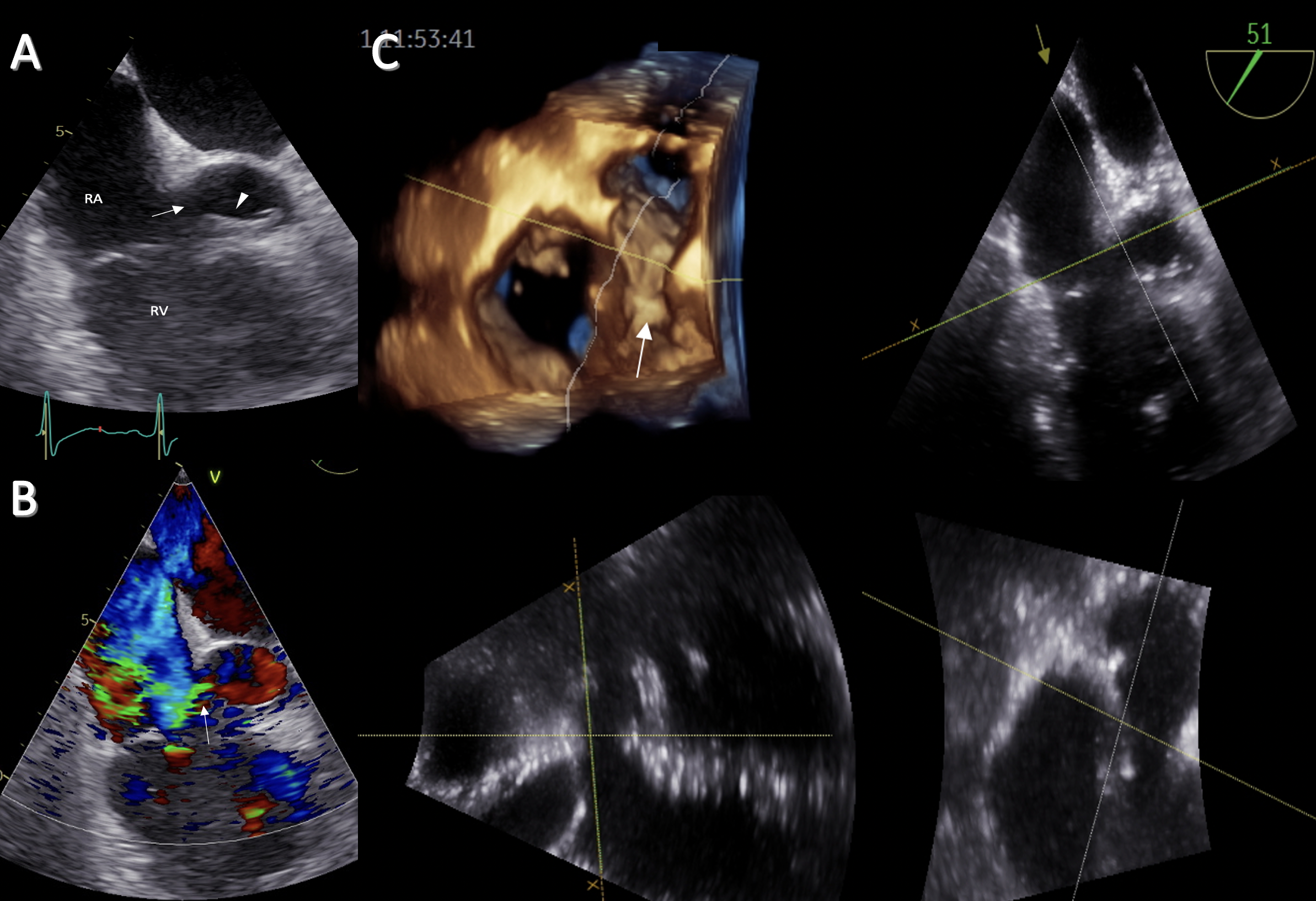
Gerbode Defect (GD) is a rare congenital heart disease that mainly affects the upper portion of the membranous septum, generating a shunt between the left ventricle and the right atrium. Even though most cases are congenital, it has also been reported acquired cases due to cardiac surgery, infective endocarditis, acute ischemic heart disease, and invasive percutaneous procedures. The diagnostic workup includes the clinical evaluation and the echocardiographic study. Here, we present the case of a 43-year-old adult patient with an incidental finding of a congenital GD in the context of acute appendicitis. Imaging plays a role in the diagnostic workup of congenital diseases; in this case, it allowed us to identify more details and the decision- making for our patient.
Downloads
References
Thurnam J. On aneurisms of the heart with cases. Medico-Chir Trans. 1838;21:187-438.9. doi: 10.1177/095952873802100114.
Gerbode F, Hultgren H, Melrose D, Osborn J. Syndrome of left ventricular-right atrial shunt; successful surgical repair of defect in five cases, with observation of bradycardia on closure. Ann Surg. 1958;148(3):433-46. doi: 10.1097/00000658-195809000-00012.
Majdoub A, Elhafidi A, Mutuale C, Boulmakoul S, Messouak M. The Gerbode Defect: About 2 Cases. World J Cardiovasc Surg. 2020;10(7):115-121. doi: 10.4236/wjcs.2020.107014.
Saker E, Bahri GN, Montalbano MJ, Johal J, Graham RA, Tardieu GG, et al. Gerbode defect: A comprehensive review of its history, anatomy, embryology, pathophysiology, diagnosis, and treatment. J Saudi Heart Assoc. 2017;29(4):283-92. doi: 10.1016/j.jsha.2017.01.006.
Eslami M, Mollazadeh R, Sattarzadeh-Badkoubeh R. Gerbode type defect after trans-septal puncture for ablation of left-sided accessory pathway. ARYA Atheroscler. 2018;14(3):139-141. doi: 10.22122/arya.v14i3.1671.
Haraf RH, Karnib M, El Amm C, Plummer S, Bocks M, Sabik EM. Gerbode defect following surgical mitral valve replacement
technique, there are more and more reports on percutaneous closure in patients with high surgical risk (5,8,9). The technical success of surgical repair is excellent, and it has low mortality rates (<3%); however, surgery in the context of acquired GD due to endocarditis or myocardial infarction could be associated with higher mortality (15-65%) (4,6).
In conclusion, despite the lack of comparative studies, patients with GD will require close clinical and echocardiographic follow-up in shorter intervals than for other septal defects. We have to bear in mind that the right atrium is the chamber with the lowest pressure, and having high pressure from the left ventricle, will cause enlargement and then increase pulmonary blood flow, which ends in pulmonary hypertension. Therefore, outpatient imaging with echocardiographic plays an important role in the close follow-up of patients with this rare condition.
Author contributions
All authors contributed equally to the idea, data collection, drafting and final approval of the manuscript.
and tricuspid valve repair: a case report. Eur Heart J-Case Rep. 2021;5(2):ytaa534. doi: 10.1093/ehjcr/ytaa534.
Sunderland N, El-Medany A, Temporal J, Pannell L, Doolub G, Nelson M, et al. The Gerbode defect: a case series. Eur Heart J Case Rep. 2021;5(2):ytaa548. doi: 10.1093/ehjcr/ytaa548.
Dhaliwal JS, Wadle MJ, Malyala R, Dwarakanath S, Hatton KW. Tricuspid valve excision complicated by postoperative Gerbode defect following recurrent infective endocarditis: A case report. Los Angeles, CA: SAGE Publications; 2021.
Ming Wang TK, Betancor J, Ho N, Rodriguez LL, Jellis CL, Griffin BP, et al. Clinical Characteristics and Multimodality Imaging–Guided Management and Outcomes of Gerbode Defects in Adults: 20-Year Experience. J Am Coll Cardiol. 2020;75(19):2520-1. doi: 10.1016/j. jacc.2020.03.010.
Kavurt AV, Ece İ, Bağrul D. Transcatheter closure of post-operative type 1 Gerbode defect by Amplatzer Duct Occluder 2. Cardiol Young. 2021;31(9):1545-7. doi: 10.1017/S1047951121002432.

Downloads
Published
Issue
Section
License
Copyright (c) 2023 The journal is headline of the first publication, then the author giving credit to the first publication.

This work is licensed under a Creative Commons Attribution 4.0 International License.


