Cafe-au-lait spots with resistant hypertension are an indicator of pheochromocytoma: a rare case report
DOI:
https://doi.org/10.47487/apcyccv.v5i2.351Palabras clave:
Cafe-au-Lait Spots, Hypertension, Pheochromocytoma, Neurofibromatosis 1Resumen
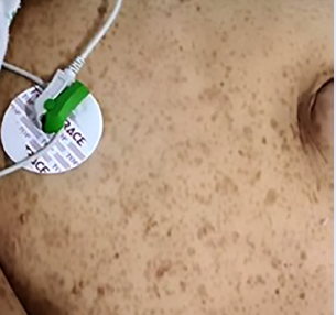
This case report is one of the rare cases of bilateral pheochromocytoma associated with neurofibromatosis type 1. The interest lies in the clinical form in which the diagnosis was revealed. We report the case of a 38-year-old woman admitted for severe hypertension resistant to triple therapy. Clinical examination revealed Cafe-au-lait spots, which are pigmented birthmarks that appear as patches on the skin with a light to dark brown colour. More than six spots are present in an estimated 95% of people diagnosed with neurofibromatosis type 1 (NF1). Abdominal computed tomography (CT) showed bilateral adrenal tumor involvement. The diagnosis of pheochromocytoma was made by measuring urinary Vanillylmandelic acid (VMA). The evolution was favorable after the excision of the tumor, with normalization of blood pressure. In conclusion: resistant hypertension with café au lait spots may indicate pheochromocytoma, especially bilateral, suggesting an underlying genetic condition like NF1, warranting systematic screening.
Descargas
Referencias
Conzo G, Pasquali D, Colantuoni V, Circelli L, Tartaglia E, Gambardella C, et al. Current concepts of pheochromocytoma. Int J Surg. 2014;12(5):469-74. doi: 10.1016/j.ijsu.2014.04.001.
Mills KT, Stefanescu A, He J. The global epidemiology of hypertension. Nat Rev Nephrol. 2020;16(4):223-237. doi: 10.1038/s41581-019-0244-2.
Siddiqui N, Daya R, Seedat F, Bulbulia S, Bayat Z. Secondary hypertension: An update on the diagnosis and localisation of a pheochromocytoma or paraganglioma. S Afr Fam Pract (2004). 2021;63(1):e1-e6. doi: 10.4102/safp.v63i1.5277.
Eya C, Lamia BH, Samira A, Zouleikha K, Imen B, Chekib K. Le phéochromocytome surrénalien bilatéral: à propos d’un cas. Pan Afr Med J. 2013;15:101. doi: 10.11604/pamj.2013.15.101.1931.
Brunaud L, Ayav A, Bresler L, Klein.M, Boissel P. Les problèmes diagnostiques du phéochromocytome. Ann Chir. 2005;130(1):267-72. doi: 10.1016/j.anchir.2005.02.008.
Raby L. Bilateral pheochromocytomas: incidental finding in a trauma patient. J Emerg Nurs. 2011;37(5):512-4. doi: 10.1016/j.jen.2011.04.018.
Garcia-Carbonero R, Matute Teresa F, Mercader-Cidoncha E, Mitjavila-Casanovas M, Robledo M, Tena I, et al. Multidisciplinary practice guidelines for the diagnosis, genetic counseling and treatment
of pheochromocytomas and paragangliomas. Clin Transl Oncol. 2021;23(10):1995-2019. doi: 10.1007/s12094-021-02622-9.
Benachour L, Haddam A.-.E.-.M, Meskine D. Un cas de phéochromocytome bilatéral avec maladie de VHL. Ann Endocrinol (Paris). 2016;77 (4): 413-414. doi: 10.1016/j.ando.2016.07.917.
Plouin PF, Gimenez-Roqueplo AP, La Batide Alanore A, Salenave S, Duclos JM. Progrès récents dans le diagnostic, l’évaluation pronostique et le traitement des phéochromocytomes. Rev Med Interne. 2000;21(12):1075-85. doi: 10.1016/s0248-8663(00)00268-x.
Pacak K, Eisenhofer G, Ahlman H, Bornstein SR, Gimenez-Roqueplo AP, Grossman AB, et al. International Symposium on Pheochromocytoma. Pheochromocytoma: recommendations for clinical practice from the First International Symposium. Nat Clin Pract Endocrinol Metab. 2007;3(2):92-102. doi: 10.1038/ncpendmet0396.
Eya C, Lamia BH, Samira A, Zouleikha K, Imen B, Chekib K, et al. Le phéochromocytome surrénalien bilatéral : à propos d’un cas. Pan Afr Med J. 2013;15:101. doi :10.11604/pamj.2013.15.101.1931.
Plouin PF, Gimenez-Roqueplo AP, La Batide Alanore A, Salenave S, Duclos JM. Progrès récents dans le diagnostic, l’évaluation pronostique et le traitement des phéochromocytomes Rev Med Interne. 2000;21(12):1075-85. doi: 10.1016/s0248-8663(00)00268.
Alface MM, Moniz P, Jesus S, Fonseca C. Pheochromocytoma: clinical review based on a rare case in adolescence. BMJ Case Rep. 2015;2015:bcr2015211184. doi: 10.1136/bcr-2015-211184.
Abid S, Rojbi I, Khelifi D, Ben Nacef I, Lakhoua Y, Mchirgui N, et al. Un phéochromocytome dans le cadre d’une Neurofibromatose type 1 révélant un syndrome de NOONAN: à propos d’un cas. Ann Endocrinol (Paris). 2020;81(4):397. doi: 10.1016/j.ando.2020.07.720
Képénékian L, Mognetti T, Lifante JC, Giraudet AL, Houzard C, Pinson S, et al. Interest of systematic screening of pheochromocytoma in patients with neurofibromatosis type 1. Eur J Endocrinol. 2016;175(4):335-44. doi: 10.1530/EJE-16-0233.
Zinnamosca L, Petramala L, Cotesta D, Marinelli C, Schina M, Cianci R, et al. Neurofibromatosis type 1 (NF1) and pheochromocytoma: prevalence, clinical and cardiovascular aspects. Arch Dermatol Res. 2011;303(5):317-25. doi: 10.1007/s00403-010-1090-z.
Boudjemaa A, Mezoued M, Bessaid K, Azzouz M. Phéochromocytome asymptomatique au cours d’une NeuroFibromatose de type 1: à propos d’un cas. Ann Endocrinol (Paris). 2021;82(5):425. doi:
1016/j.ando.2021.08.494.
BeRebai S, Debbabi W, Kammoun F, Chermit S, Marzouk H, Kharrat I, et al. Neurofibromatose découverte en post opératoire suite à une rupture spontanée d’un phéochromocytome méconnu.
Ann Endocrinol (Paris). 2021;82(5):439-440. doi: 10.1016/j.ando.2021.08.539.
Belhimer F, Fedala S. Maladie de Von Reklinhausen et phéochromocytome. Ann Endocrinol (Paris). 2018;79(4):392. doi : 10.1016/j.ando.2018.06.635.
Képénékian L, Mognetti T, Lifante JC, Giraud, Houzard C, Pinson S, et al. Interest of systematic screening of pheochromocytoma in patients with neurofibromatosis type 1. Eur J Endocrinol. 2016;175(4):335-44. doi: 10.1530/EJE-16-0233.
Neumann HPH, Tsoy U, Bancos I, Amodru V, Walz MK, Tirosh A, et al. International Bilateral-Pheochromocytoma-Registry Group. Comparison of Pheochromocytoma-Specific Morbidity and Mortality Among Adults with Bilateral Pheochromocytomas Undergoing Total Adrenalectomy vs Cortical-Sparing Adrenalectomy. JAMA Netw Open. 2019;2(8):e198898. doi: 10.1001/jamanetworkopen.2019.8898.
Boaz RJ, Ramakant P, Ebenazer A, Pai R, Rajaratnam S, Abraham D, et al. Role of cortical sparing adrenalectomy and novel variant of mutation in patient with von Hippel-Lindau disease. Indian J Endocrinol Metab. 2011;15 Suppl 4(Suppl4):S402-5. doi: 10.4103/2230-8210.86987.
Alesina PF, Knyazeva P, Hinrichs J, Walz MK. Tailored Approach in Adrenal Surgery: Retroperitoneoscopic Partial Adrenalectomy. Front Endocrinol (Lausanne). 2022;13:855326. doi: 10.3389/fendo.2022.855326.
Descargas
Publicado
Número
Sección
Licencia
Derechos de autor 2024 La revista es titular de la primera publicación, luego el autor dando crédito a la primera publicación.

Esta obra está bajo una licencia internacional Creative Commons Atribución 4.0.