Prevalencia de anomalías coronarias detectadas por tomografía computarizada en el Instituto Nacional Cardiovascular - INCOR
DOI:
https://doi.org/10.47487/apcyccv.v3i3.233Palabras clave:
Arterias Coronarias, Angiografía por Tomografía Computarizada, Malformaciones de los Vasos Coronarios, PerúResumen
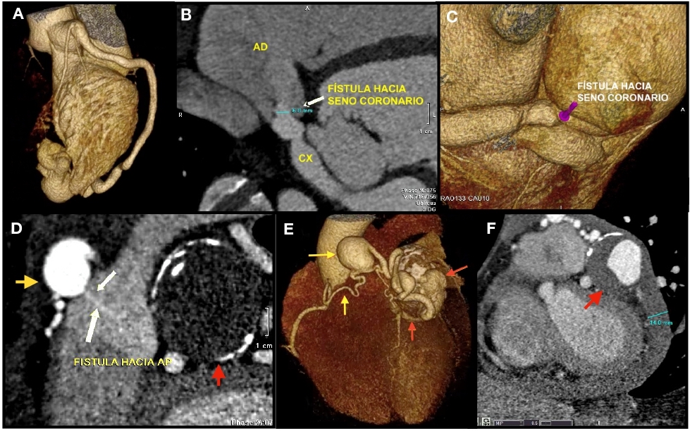
Objetivo: determinar la prevalencia de anomalías coronarias (AC) en pacientes evaluados por tomografía computarizada (TC) de 64 detectores en el Instituto Nacional Cardiovascular en el Perú entre los años 2016 a 2020. Materiales y métodos: estudio observacional retrospectivo, en el cual se revisaron las TC de arterias coronarias de 1486 pacientes, realizadas en un tomógrafo de 64 filas de detectores, en busca de anomalías coronarias. Resultados: la prevalencia de AC detectada por TC fue de 4,71% (70 casos) de ellos 64,3% varones. Las anomalías de origen fueron las más frecuentes, de ellas el nacimiento de una arteria coronaria desde el seno coronariano opuesto fue la más común (48,6%), siendo la coronaria derecha la principal arteria anómala (31%), y el principal trayecto fue el interarterial (31%). El origen anómalo del tronco coronario izquierdo desde la arteria pulmonar se encontró en cinco pacientes. Entre las anomalías de la anatomía arterial coronaria intrínseca, la principal fue la doble arteria descendente anterior (10%). Las fístulas coronarias representaron el 11,4% de casos. Conclusiones: la prevalencia de AC detectadas por TC de 64 detectores en un instituto del Perú fue de 4,71%. La principal anomalía coronaria fue el origen de la arteria coronaria derecha desde el seno coronariano izquierdo con trayecto interarterial.
Descargas
Referencias
Angelini P. Normal and anomalous coronary arteries: definitions and classification. Am Heart J. 1989;117(2):418-34. doi: 10.1016/0002- 8703(89)90789-8.
Angelini P. Coronary Artery Anomalies. An entity in Search of an Identity. Circulation. 2007;115:1296-1305. doi: 10.1161/ CIRCULATIONAHA.106.618082.
Villa AD, Sammut E, Nair A, Rajani R, Bonamini R, Chiribiri A. Coronary artery anomalies overview: The normal and the abnormal. World J Radiol. 2016;8(6):537-55. doi: 10.4329/wjr.v8.i6.537.
Ghadri JR, Kazakauskaite E, Braunschweig S, Burger IA, Frank M, Fiechter M, et al. Congenital coronary anomalies detected by coronary computed tomography compared to invasive coronary angiography. BMC Cardiovasc Disord. 2014;14:81. doi: 10.1186/1471-2261-14-81.
Budoff MJ, Ahmed V, Gul KM, Mao SS, Gopal A. Coronary anomalies by cardiac computed tomographic angiography. Clin Cardiol. 2006;29(11):489- 93. doi: 10.1002/clc.4960291104.
Cheezum MK, Ghoshhajra B, Bittencourt MS, Hulten EA, Bhatt A, Mousavi N, et al. Anomalous origin of the coronary artery arising from the opposite sinus: prevalence and outcomes in patients undergoing coronary CTA. Eur Heart J Cardiovasc Imaging. 2017;18(2):224-235. doi: 10.1093/ehjci/jev323.
Stout KK, Daniels CJ, Aboulhosn JA, Bozkurt B, Broberg CS, Colman JM, et al. 2018 AHA/ACC Guideline for the Management of Adults With Congenital Heart Disease: Executive Summary: A Report of the American College of Cardiology/American Heart Association Task Force on Clinical Practice Guidelines. J Am Coll Cardiol. 2019;73(12):1494-1563. doi: 10.1016/j.jacc.2018.08.1028.
Di Salvo G, Miller O, Babu Narayan S, Li W, Budts W, Valsangiacomo Buechel ER, et al.; 2016–2018 EACVI Scientific Documents Committee. Imaging the adult with congenital heart disease: a multimodality imaging approach-position paper from the EACVI. Eur Heart J Cardiovasc Imaging. 2018;19(10):1077-1098. doi: 10.1093/ehjci/jey102.
L i p t o n M J , B a r r y W H , O b r e z I , S i l v e r m a n J F, W e x l e r L . I s o l a t e d s i n g l e c o r o n a r y artery: diagnosis, angiographic classification, and clinical significance. Radiology. 1979;130(1):39-47. doi: 10.1148/130.1.39.
Spindola-Franco H, Grose R, Solomon N. Dual left anterior descending coronary artery: angiographic description of important variants and surgical implications. Am Heart J. 1983;105(3):445-455. doi: 10.1016/0002- 8703(83)90363-0.
Maron BJ, Doerer JJ, Haas TS, Tierney DM, Mueller FO. Sudden deaths in young competitive athletes: analysis of 1866 deaths in the United States, 1980-2006. Circulation. 2009;119(8):1085-92. doi: 10.1161/ CIRCULATIONAHA.108.804617
Pérez-Pomares JM, de la Pompa JL, Franco D, Henderson D, Ho SY, Houyel L, et al. Congenital coronary artery anomalies: a bridge from embryology to anatomy and pathophysiology--a position statement of the development, anatomy, and pathology ESC Working Group. Cardiovasc Res. 2016;109(2):204-16. doi: 10.1093/cvr/cvv251.
Sundaram B, Kreml R, Patel S. Imaging of coronary artery anomalies. Radiol Clin North Am. 2010;48(4):711-727. doi: 10.1016/j.rcl.2010.04.006.
Angelini P, Velasco JA, Flamm S. Coronary anomalies: incidence, pathophysiology, and clinical relevance. Circulation. 2002;105(20):2449-54. doi: 10.1161/01.cir.0000016175.49835.57.
Sirasapalli CN, Christopher J, Ravilla V. Prevalence and spectrum of coronary artery anomalies in 8021 patients: A single center study in South India. Indian Heart J. 2018;70(6):852-856. doi: 10.1016/j.ihj.2018.01.035.
Namgung J, Kim JA. The prevalence of coronary anomalies in a single center of Korea: origination, course, and termination anomalies of aberrant coronary arteries detected by ECG-gated cardiac MDCT. BMC Cardiovasc Disord. 2014;14:48. doi: 10.1186/1471-2261-14-48.
Gräni C, Benz DC, Schmied C, Vontobel J, Possner M, Clerc OF, et al. Prevalence and characteristics of coronary artery anomalies detected by coronary computed tomography angiography in 5634 consecutive patients in a single centre in Switzerland. Swiss Med Wkly. 2016;146:w14294. doi: 10.4414/smw.2016.14294.
Schmitt R, Froehner S, Brunn J, Wagner M, Brunner H, Cherevatyy O, et al. Congenital anomalies of the coronary arteries: imaging with contrast-enhanced, multidetector computed tomography. Eur Radiol. 2005;15(6):1110-21. doi: 10.1007/s00330-005-2707-z.
Shabestari AA, Akhlaghpoor S, Tayebivaljozi R, Fattahi Masrour F. Prevalence of Congenital Coronary Artery Anomalies and Variants in 2697 Consecutive Patients Using 64-Detector Row Coronary CTAngiography. Iran J Radiol. 2012;9(3):111-21. doi: 10.5812/iranjradiol.8070.
Cheezum MK, Liberthson RR, Shah NR, Villines TC, O’Gara PT, Landzberg MJ, Blankstein R. Anomalous Aortic Origin of a Coronary Artery From the Inappropriate Sinus of Valsalva. J Am Coll Cardiol. 2017;69(12):1592-1608. doi: 10.1016/j.jacc.2017.01.031.
Yamanaka O, Hobbs RE. Coronary artery anomalies in 126,595 patients undergoing coronary arteriography. Cathet Cardiovasc Diagn. 1990;21(1):28- 40. doi: 10.1002/ccd.1810210110.
Frescura C, Basso C, Thiene G, Corrado D, Pennelli T, Angelini A, et al. Anomalous origin of coronary arteries and risk of sudden death: a study based on an autopsy population of congenital heart disease. Hum Pathol. 1998;29(7):689-95. doi: 10.1016/s0046-8177(98)90277-5.
Smith JC. Review of single coronary artery with report of 2 cases. Circulation 1950;1:1168-75. doi: 10.1161/01.cir.1.5.1168.
Michalowska AM, Tyczynski P, Pregowski J, Skowronski J, Mintz GS, Kepka C, et al. Prevalence and Anatomic Characteristics of Single Coronary Artery Diagnosed by Computed Tomography Angiography. Am J Cardiol. 2019;124(6):939-946. doi: 10.1016/j.amjcard.2019.06.012.
Desmet W, Vanhaecke J, Vrolix M, Van de Werf F, Piessens J, Willems J, de Geest H. Isolated single coronary artery: a review of 50,000 consecutive coronary angiographies. Eur Heart J. 1992;13(12):1637-40. doi: 10.1093/ oxfordjournals.eurheartj.a060117.
Şeker M. Prevalence and morphologic features of dual left anterior descending artery subtypes in coronary CT angiography. Radiol Med. 2020;125(3):247-256. doi: 10.1007/s11547-019-01124-7.
Barbaryan A, Addai T, Kola M, Raqeem MW, Barsamyan S, Mirrakhimov AE. A Combination of Two Rare Coronary Anomalies Makes It Even Rarer: Right Sided Single Coronary Artery with Dual Left Anterior Descending Artery. Case Rep Cardiol. 2016;2016:4905941. doi: 10.1155/2016/4905941.
Chen YF, Chien TM, Chen CW, Lin CC, Lee CS. Double right coronary artery or split right coronary artery? Int J Cardiol. 2012;154(3):243-5. doi: 10.1016/j. ijcard.2011.10.053.
Kunimasa T, Sato Y, Ichikawa M, Ito S, Takagi T, Lee T, et al. MDCT detection of double right coronary artery arising from a single ostium in the right sinus of Valsalva: report of 2 cases. Int J Cardiol. 2007;115(2):239-41. doi: 10.1016/j.ijcard.2006.01.060.
Kodama-Takahashi K, Suzuki J, Watanabe A, Ohtsuka T, Hashida H, Ikeda S, Kuwahara T, Hara Y, Shigematsu Y, Hamada M, Hiwada K. Ischemia in the territory of the first major septal perforator branch anomalously originating from the first diagonal branch leads to a transient leftward shift of the QRS axis in the frontal plane: a case report. Circ J. 2003;67(10):885-8. doi: 10.1253/circj.67.885.
Verna E, Santarone M, Boscarini M, Ghezzi I, Repetto S. Unusual origin and course of the first septal branch of the left coronary artery: angiographic recognition. Cardiovasc Intervent Radiol. 1988;11(3):146-9. doi: 10.1007/ BF02577106.
Yun G, Nam T, Chun E. Coronary Artery Fistulas: Pathophysiology, Imaging Findings, and Management. Radiographics. 2018;38(7):688-703. doi: 10.1148/rg.2018170158.
Vavuranakis M, Bush CA, Boudoulas H. Coronary artery fistulas in adults: incidence, angiographic characteristics, natural history. Cathet Cardiovasc Diagn. 1995;35(2):116-20. doi: 10.1002/ccd.1810350207.
Lim JJ, Jung JI, Lee BY, Lee HG. Prevalence and types of coronary artery fistulas detected with coronary CT angiography. AJR Am J Roentgenol. 2014;203(3):W237-43. doi: 10.2214/AJR.13.11613.
Publicado
Número
Sección
Licencia
Derechos de autor 2022 La revista es titular de la primera publicación, luego el autor dando crédito a la primera publicación.

Esta obra está bajo una licencia internacional Creative Commons Atribución 4.0.