Rotura de aislante de electrodo de marcapasos por atrapamiento subclavio. Reporte de caso
DOI:
https://doi.org/10.47487/apcyccv.v3i2.210Palabras clave:
marcapasos, Falla del electrodo, Vena subclavia, Vena AxilarResumen
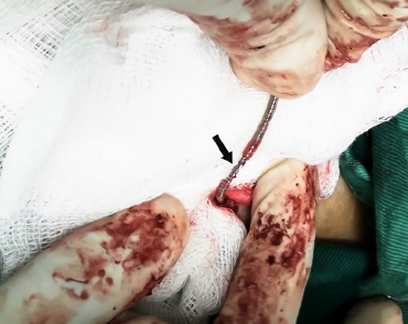
Se presenta el caso de un paciente varón de 82 años con antecedente de implante de marcapasos unicameral hace 7 años, por bloqueo auriculoventricular de tercer grado, que experimenta presíncopes. En la telemetría se encuentró alerta de posible daño de cable electrodo y entrada de ruido ventricular que coincide con falla de captura, por lo que se programó reemplazo de todo el sistema de estimulación. Durante la intervención se observó rotura del aislante del electrodo. El abordaje subclavio puede ocasionar daño en el electrodo por atrapamiento en los tejidos blandos que atraviesa antes de ingresar al sistema venoso y estiramiento iterativo. La rotura del aislante se puede manifestar como pérdida de captura y presencia de ruido en el electrocardiograma, caída de la impedancia del cable e imágenes radiográficas sugerentes. El abordaje axilar es una opción segura y eficaz, por lo que se recomienda para el implante de cables transvenosos.
Descargas
Referencias
Bass D, Stephenson K, Gyi R, Zeltser R, Makaryus A. Subclavian Crush Syndrome: A Rare Cause of Symptomatic Bradycardia in a Patient with a Permanent Pacemaker. J Cardiovasc Dis Diagn. 2017;5:267. doi: 10.4172/2329- 9517.1000267.
Espinosa FR. Subclavian crush syndrome. Complete rupture of the pacemaker electrode. Minutes Med Group Angeles. 2021;19(2):294-295. doi: 10.35366/100460.
Magney JE, Flynn DM, Parsons JA, Staplin DH, Chin-Purcell MV, Milstein S, et al. Anatomical mechanisms explaining damage to pacemaker leads, defibrillator leads, and failure of central venous catheters adjacent to the sternoclavicular joint. Pacing Clin Electrophysiol. 1993;16(3 Pt 1):445-57. doi: 10.1111/j.1540-8159.1993.tb01607.x.
Luo GH, Li WJ, Zhong SZ, Li ZH, Fang J. Modification of the right subclavian vein catheterization and its anatomic basis and techniques. Chin Med J. 2005;118(8):645-53.
Kotter J, Lolay G, Charnigo R, Leung S, McKibbin C, Sousa M, et al. Predictors, morbidity, and costs associated with pneumothorax during electronic cardiac device implantation. Pacing Clin Electrophysiol. 2016;39(9):985-91. doi: 10.1111/pace.12901.
Kirkfeldt RE, Johansen JB, Nohr EA, Moller M, Arnsbo P, Nielsen JC. Pneumothorax in cardiac pacing: a population-based cohort study of 28,860 Danish patients. Europace. 2012;14(8):1132-8. doi: 10.1093/europace/eus054.
Benz AP, Vamos M, Erath JW, Hohnloser SH. Cephalic vs. subclavian lead implantation in cardiac implantable electronic devices: a systematic review and meta-analysis. Europace. 2019;21(1):121-129. doi: 10.1093/europace/euy165.
Chan NY, Kwong NP, Cheong AP. Venous access and long-term pacemaker lead failure: comparing contrast-guided axillary vein puncture with subclavian puncture and cephalic cutdown. Europace. 2017;19(7):1193-1197. doi: 10.1093/europace/euw147.
Roelke M, O’Nunain SS, Osswald S, Garan H, Harthorne JW, Ruskin JN. Subclavian crush syndrome complicating transvenous cardioverter defibrillator systems. Pacing Clin Electrophysiol. 1995;18(5 Pt 1):973-9. doi: 10.1111/j.1540-8159.1995.tb04737.x.
Swerdlow CD, Koneru JN, Gunderson B, Kroll MW, Ploux S, Ellenbogen KA. Impedance in the diagnosis of lead malfunction. Circ Arrhythm Electrophysiol. 2020 Feb;13(2):e008092. doi: 10.1161/CIRCEP.119.008092.
Ellenbogen KA, Gunderson BD, Stromberg KD, Swerdlow CD. Performance of lead integrity alert to assist in the clinical diagnosis of implantable car-dioverter defibrillator lead failures: analysis of different implantable cardio-verter defibrillator leads. Circ Arrhythm Electrophysiol. 2013;6(6):1169-77. doi: 10.1161/CIRCEP.113.000744.
Safavi-Naeini P, Saeed M. Pacemaker troubleshooting: common clinical scenarios. Texas Heart Inst J. 2016;43(5):415-418. doi: 10.14503/THIJ-16-5918.
Sabbagh E, Abdelfattah T, Karim MM, Farah A, Grubb B, Karim S. Causes of Failure to Capture in Pacemakers and Implantable Cardioverter-defibrillators. J Innov Card Rhythm Manag. 2020;11(2):4013-4017. doi: 10.19102/icrm.2020.110207.
Hauser RG, Hayes DL, Kallinen LM, Cannom DS, Epstein AE, Almquist AK, et al. Clinical experience with pacemaker pulse generators and transvenous leads: an 8-year prospective multicenter study. Heart Rhythm. 2007;4(2):154-60. doi: 10.1016/j.hrthm.2006.10.009.
Kowalski M, Ellenbogen KA, Wood MA, Friedman PL. Implantable cardiac defibrillator lead failure or myopotential oversensing? An approach to the diagnosis of noise on lead electrograms. Europace. 2008;10(8):914-7. doi: 10.1093/europace/eun167.
Ajit Deshpande S, Udyavar A. A case report of lead dysfunction presenting as high ventricular premature complex burden. Indian Pacing Electrophysiol J. 2022;22(1):34-37. doi:10.1016/j.ipej.2021.10.003.
Jimenez-Diaz J, Higuera-Sobrino F, Piqueras-Flores J, Perez-Diaz P, Gonzalez-Marin MA. Fluoroscopy-guided axillary vein access vs cephalic vein access in pacemaker and defibrillator implantation: randomized clinical trial of efficacy and safety. J Cardiovasc Electrophysiol. 2019;30(9):1588-1593. doi: 10.1111/jce.14060.
Yang F, Kulbak G. A new trick to a routine procedure: taking the fear out of the axillary vein stick using the 35° caudal view. Europace. 2015;17(7):1157-60. doi: 10.1093/europace/euv066.
Liccardo M, Nocerino P, Gaia S, Ciardiello C. Efficacy of ultrasound-guided axillary/subclavian venous approaches for pacemaker and defibrillator lead implantation: a randomized study. J Interv Card Electrophysiol. 2018;51(2):153-160. doi: 10.1007/s10840-018-0313-7.
Burri H, Starck C, Auricchio A, Biffi M, Burri M, D’Avila ALR, et al. EHRA expert consensus statement and practical guide on optimal implantation technique for conventional pacemakers and implantable cardioverter-defibrillators: endorsed by the Heart Rhythm Society (HRS), the Asia Pacific Heart Rhythm Society (APHRS), and the Latin-American Heart Rhythm Society (LAHRS). Europace. 2021;23(7):983-1008. doi: 10.1093/europace/euaa367.
Publicado
Número
Sección
Licencia
Derechos de autor 2022 La revista es titular de la primera publicación, luego el autor dando crédito a la primera publicación.

Esta obra está bajo una licencia internacional Creative Commons Atribución 4.0.